


Description
A corneal ulcer occurs when the protective surface layer of the eye’s cornea is lost or damaged. Corneal ulcers range from superficial abrasions and small circular lesions to deep craters. The deeper layers of the cornea are exposed and become prone to infection and injury. These deeper layers contain many nerves, and irritation of these nerves is very painful. Ulcers may be complicated by secondary bacterial infections.
Causes
There are many causes of corneal ulceration, including:
• Trauma from cat scratches or other foreign objects
• Abnormalities of the eyelids including entropion (eyelids rolled inward) or abnormal ingrown eyelashes
• Decreased blinking due to neurologic problems or from sedation and general anesthesia
• Exposure to irritants such as chemicals, soaps, or smoke
• Infections with bacteria, viruses, or fungal agents
• Chronic dry eyes from poor tear production
• Corneal infiltrates such as calcium, cholesterol, or edema
• Brachycephalic breeds of dogs or cats with flat faces and prominent eyes are very prone to corneal ulcers because their eyes protrude beyond the eyelids.
Clinical Signs
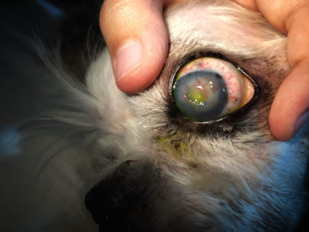
Pain is a hallmark sign of corneal injuries and ulceration. A pet with a painful eye may have increased tearing, squinting, blinking, and pawing at the eye. The animal may also be quiet and withdrawn. The eye is usually red and thick eye discharge may develop with infection. The cornea may be cloudy or have visible irregularities. Other signs, such as swelling of the eyelid, inward rolling of the eyelid, protrusion of the third eyelid, or bruising around the head, may occur depending on the cause of the ulceration. Signs of an upper respiratory tract infection may be noted in cases of herpes virus infection in cats or distemper virus infection in dogs.
 Diagnostic Tests
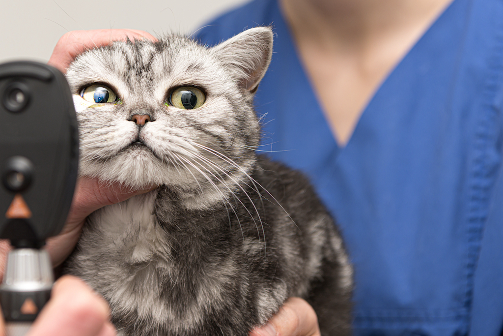
A veterinarian diagnosis a corneal ulcer by performing an ophthalmic exam.
Fluorescein staining of the cornea is used to confirm the presence of a corneal ulcer. The stain adheres to deeper corneal layers that are exposed when the corneal surface is damaged. The damaged area if cornea is visible as apple green under blue light. Other ocular tests such as a Schirmer tear test, testing of reflexes, examination of the eyelids and interior of the eye, and glaucoma testing may also be indicated. If a severe bacterial infection is suspected a culture may need to be submitted.
TREATMENT AND FOLLOW-UP
 Treatment Options
Most corneal ulcers in dogs and cats can be treated on an outpatient basis by a general veterinarian.
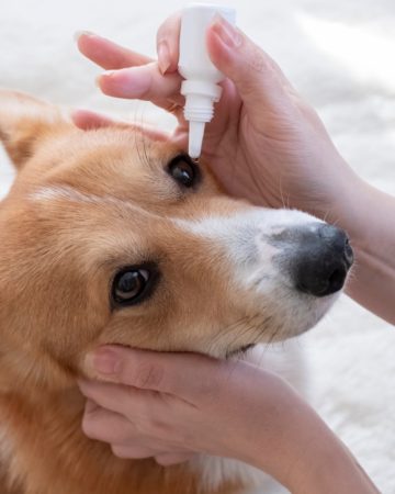
Medications and treatments prescribed for corneal ulcers:
• Topical antibiotics drops or ointment administered directly to the eye to treat active infections and/or to prevent infections.
• If the ulcer is deep and there is concern about infection within the eye, then oral antibiotics may also be prescribed.
• Topical atropine (pupil dilator) may be administered as an ointment or drop directly to the eye.
• Oral nonsteroidal anti-inflammatory drugs may be considered for marked inflammation and discomfort.
• Administration of the pet’s own blood serum as eye drops to provide nutrients to encourage healing.
• Elizabethan cone collar to prevent self-trauma.
• Debridement of the ulcer with cotton swab to remove dead tissue
Referral to a veterinary ophthalmologist may be recommended if the ulcer is deep or not healing. An ophthalmologist can offer application of soft contact bandage lenses or placement of a third eyelid flap to protect the cornea. An ophthalmologist can also offer treatments for underlying conditions such as entropion correction, removal of foreign materials, or abnormal eye lashes.
 Follow-up Care
Recheck exams are needed to assess healing and response to treatment. Visit frequency can range from every 24-48 hours to every 7-14 days, depending on the severity of the corneal ulcer. Fluorescein staining of the cornea is performed at most visits to highlight the ulcer and to determine when it has healed. Brachycephalic breeds such as boxers, bull dogs, and Persians may need more intensive follow-up.
Prognosis
Most superficial corneal ulcers and abrasions caused by trauma respond to treatment quickly and heal with minimal scarring. Pets that have underlying chronic conditions such as entropion or ingrown eye lashes may need to have minor procedures to correct the abnormality. Chronic conditions, such as dry eye, may need lifelong treatment with eye drops to prevent future corneal ulcers. Deep ulcers and ulcers that are infected are more difficult to treat and may leave the cornea scarred and pigmentated resulting in decreased vision. In rare instances corneal ulcers that that do not receive treatment and become infected can result in perforations of the eye and loss of vision, and in severe cases even loss of the eye.

Recent Comments