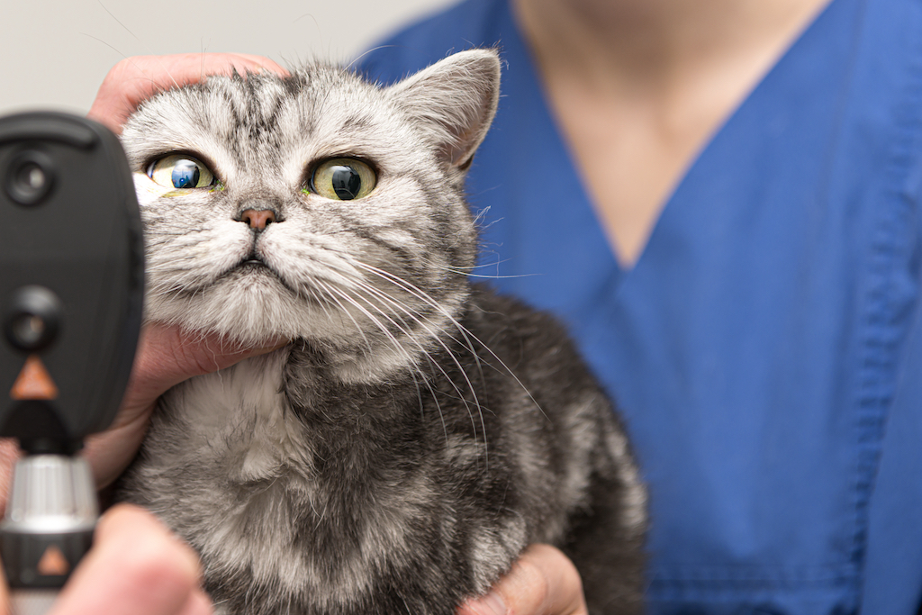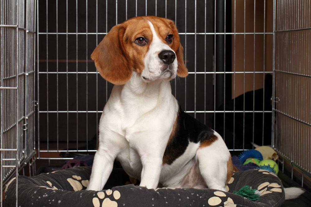
Keratoconjunctivitis Sicca (Dry Eye)

Keratoconjunctivitis sicca (KCS) is the medical term for dry eye

Causes of KCS

Dry eye in pets is commonly caused by an immune-mediated inflammation of the tear glands.  Chronic inflammation damages the gland and causes decreased production of the essential watery component of tears. Tears play an important role in maintaining the health of the ocular surface, protecting the eye from infection and irritants.

KCS is a very common canine eye disease with a reported incidence of 1% to 2% of dogs in North America. By contrast, the incidence of dry eye in cats is very low. The disease most likely has a genetic component, as it is more prevalent in some breeds including the Cocker spaniel, English bulldog, Lhasa Apso, Shih Tzu, West Highland White Terrier, Schnauzer, Pug, Cavalier King Charles Spaniel, among others. Dogs that have a history of a prolapsed gland of the third eyelid (cherry eye) may also be pre-disposed to dry eye.


Clinical Signs of KCS

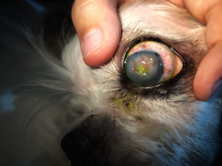
When there is a decrease in tear production, drying and inflammation of the surface tissues of the eye occur. The conjunctiva and cornea become red and inflamed. Thick yellow to green ocular discharge may build up around the eye. Secondary corneal ulceration commonly occurs. The cornea may become pigmented and scarred from chronic inflammation.  Brachycephalic dogs with large protruding eyes, including pugs or bull dogs, may be more severely impacted by the disease.

Common Symptoms of Dry Eye


Squinting or holding the eyes closed

  • Rubbing the face and eyes 
  • Red or inflamed eyes
  • Thick goopy ocular discharge near the eyelids
  • Corneas appear dull and dry
  • Secondary conjunctivitis, infections, or ulcers
  • Dark pigment on the eye surface
  • Prominent blood vessels on the eyes

Diagnosis of KCS

KCS may tentatively be suspected based on history and clinical signs. On physical exam a veterinarian may find thick ocular discharge, a cornea that appears dry or ulcerated, and corneal pigmentation.

KCS diagnosis is confirmed by measurement of tear production using the Schirmer tear test. Dogs that produce less than 10mm of tears over a minute on a Schirmer tear test have insufficient tear production and are likely suffering from KCS. The tear test may be performed on more than one occasion, because the results can vary somewhat. Other causes of conjunctivitis, keratitis, and thick discharge are ruled out with a thorough eye examination.

Treatment of KCS

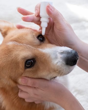
The main goal of treatment is to control immune-mediated KCS by stimulating natural production of the tear film’s aqueous component. Topical cyclosporine or tacrolimus are the cornerstone of KCS immunomodulating therapy.

Cyclosporine is FDA approved and commercially available as an ointment. An additional medication, Tacrolimus, is available as a treatment from compounding pharmacies. Either of these drugs is administered 1-3 times daily, often life long. Tear production may increase within 3-4 weeks, but it can take as long as 12 weeks to see a response. 


In addition to immunomodulators, keeping the eyes well lubricated protects the cornea and increases patient comfort. A variety of products, such as Optixcare eye lube, are available for animal eyes. Topical antibiotics such as Neo/Poly/Bac ointment may also be administered to treat secondary corneal ulcers, infections, and conjunctivitis. 


Prognosis of KCS

KCS is a lifelong and chronic disease that may be controllable but is not curable. Regular diligent administration of eye medications by the pet owner and follow-up progress exams with a your veterinarian are key to managing dry eye. Eyes that respond to tear stimulant medications have a good prognosis. Medications need to be administered daily, response to therapy monitored, and treatment adjustments made to maintain the health and comfort of the eye. 

Unfortunately, some KCS patients may have a guarded or poor prognosis. Not all patients respond to the available medications.  Some patients may need referral to a veterinary ophthalmologist and numerous medications used to manage their dry eye. If untreated, KCS has a poor prognosis. Untreated dry eye can cause chronic pain, compromise a pet’s quality of life, and may cause permanent damage to eyesight.

Intervertebral Disk Disease of Dogs

Intervertebral disc disease (IVDD) is a degenerative disease process of the spine that can result in back pain and loss of mobility.

What causes IVDD

The intervertebral discs are the fibrous cartilage structure between the boney vertebrae of the spine. An intervertebral disc is composed of two parts with a structure similar to a jelly donut. (IVDD) is a degenerative disease process of the spine that can result in back pain and loss of mobility.

What causes IVDD

The intervertebral discs are the fibrous cartilage structure between the boney vertebrae of the spine. An intervertebral disc is composed of two parts with a structure similar to a jelly donut. The outer covering of the disk is a thick, tough, and fibrous shell called the annulus fibrosis. The annulus fibrosis is the outer chewy dough of the donut. The inner squishy middle of the disk is called the nucleus pulposus. The nucleus pulposus is the jelly center of the doughnut.

Intervertebral Disc Disease (IVDD) is an age-related degenerative process that affects the spinal cord of dogs. Degenerative disc disease causes deterioration of the outer annulus fibrosus, which can result in a bulging or herniation of the disc. The spinal cord is encased within the boney canal of the vertebrae and cannot move away from the pressure of bulging or herniating discs. Compression of the spinal cord results in pain and loss of neurologic function.
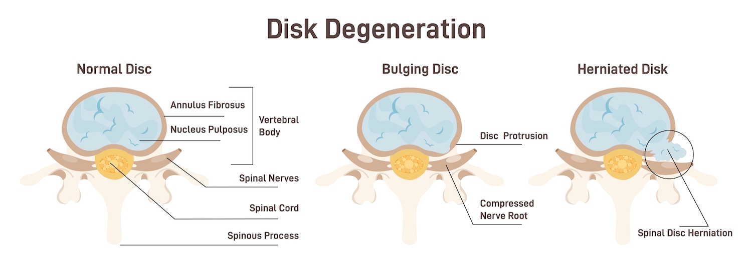
There are two types of intervertebral disc disease.

Type 1 – IVDD type 1 occurs in small dogs that have longer backs and short legs including dachshunds, corgis, basset hounds, Lhasa Apsos, Chihuahuas, toy poodles, French bulldogs, beagles, and Shih Tzus. The center of the disc herniates through a tear in the outer part of the disc, similar to jam bursting out of the donut. This is referred to as a herniated disk. A herniated disc causes acute compression of the spinal cord and can result in sudden onset of pain, difficulty walking, and paralysis.

Type 2 – IVDD type 2 occurs in large dogs such as labradors, Dobermans Pinschers, and German Shepherds. The fibrous outer layer of the annulus fibrosus starts to deteriorate allowing both the outer and inner parts of the disc to bulge out and compress the spinal cord. This is similar to a bulging donut. IVDD type 2 has a slow chronic onset and may be less painful. There may not be a particular moment or action that can be identified as having caused the damage.

Symptoms of IVDD

Intervertebral Disc Disease symptoms depend upon which part of the spine is affected and how severely the spinal cord is compressed. Symptoms of IVDD may appear suddenly or come on gradually.
Symptoms of Intervertebral Disc Disease (IVDD) may include:
• Holding the neck low or unwillingness to lift the head
• Hunched back and stiff appearance
• Avoidance of normal activities such as climbing stairs
• Shivering, panting, or crying due to pain
• Reluctance to move
• Unsteadiness or weakness in limbs
• Uncoordinated movements and difficulty walking
• Knuckling or dragging of paws
• Paralysis of limbs
• Urinary or fecal incontinence

If a dog is displaying any of the above symptoms, seek veterinary care as soon as possible. IVDD can be very painful for dogs and early treatment is essential for preventing the condition from becoming more severe. Delayed treatment may cause irreversible damage to the spine.

Diagnosis of IVDD

Intervertebral disk disease is diagnosed based on clinical signs, exam findings, and imaging.

Physical Exam – A complete physical exam that includes neurologic and orthopedic evaluation should be performed by a veterinarian. Physical exam findings can help pinpoint the pain location along the spine.

Radiographs — Radiographs can identify evidence of disc degeneration, calcification, and narrowing of disk spaces. Other bone lesions such as cancers or infections that may cause similar symptoms to IVDD can also be identified on radiographs. However, because the spinal cord does not appear on X-rays, radiographs cannot definitively diagnose IVDD.

Magnetic resonance imaging (MRI) — A definitive diagnosis of IVDD is usually achieved with advanced imaging of the spine. An MRI can best diagnose IVDD because the image provides details of all the spinal structures and identifies the degree of spinal cord compression and the side affected.

Treatment for IVDD

The treatment for Intervertebral Disc Disease needs to begin as early as possible to achieve good treatment outcomes. Your dog to the vet for a full examination if you spot signs of IVDD. Delays in treatment could lead to irreversible spinal damage.

Anti-Inflammatory Medications

If a patient is diagnosed with a mild to moderate IVDD, treatment may include non-steroidal anti-inflammatory medications (NSAIDS) such as Carprofen, Deracoxib, or Meloxicam. Some patients may respond better to steroids such as prednisone or methylprednisolone to help reduce pain and swelling.

Pain medications and Muscle relaxers

Patients with moderate IVDD symptoms may benefit from Gabapentin, a medication that targets nerve-related pain. Many patients may be more comfortable if also prescribed muscle relaxers such as methocarbamol. Occasionally, patients with severe pain may be prescribed opioids such as codeine.

Activity Restriction

Patients suffering from IVDD should have strict rest imposed the first 4 weeks. Rest helps minimize the strain on the spine, intervertebral discs, and surrounding musculature.
Patients should be confined to a small well-padded area, such as a kennel, for the first 2 weeks to impose bed rest. Absolutely no running, jumping, climbing, or playing should be allowed. The patient should be carried or escorted outside on a short leash for supervised bathroom breaks.

The patient can begin to return to some minor physical activity gradually over the next two weeks. Start with brief leash walks multiple times a day. Patients should continue to be confined to a small area when they are not supervised. Activities such as climbing, jumping, and running should still be restricted.

Acupuncture & Physical Therapy

Several studies have shown that IVDD patients that have acupuncture and physical therapy added to their treatment plan recover faster and better. Stimulation of acupuncture points help the body release endorphins, norepinephrine, and anti-inflammatory mediators that relieve pain and help the body to heal itself naturally. Physical therapy helps IVDD patients to gain strength and mobility. Targeted exercises help to build muscle mass, regain stability, and recover range of motion, allowing patients to recover quality of life sooner.

Surgery

Surgery is typically recommended for dogs suffering from more severe cases of Intervertebral Disc Disease. Patients that are experiencing paralysis may need surgical decompression of the spinal cord, when rest and medication are not sufficient to reduce pain and symptoms. During surgery the disc material which is pressing onto the spinal cord and causing the IVDD symptoms is removed.

Prognosis of IVDD

The prognosis for dogs with IVDD depends on several factors including the severity of the clinical signs, the location of the spinal cord compression, how quickly the patient receives treatment, and initial response to therapy.
Patients that have mild symptoms and receive veterinary care within 24 hours will likely improve with noninvasive care including medication and rest. These patients have a good prognosis. However, IVDD is a degenerative disease, symptoms can reoccur and get worse over time. Patients that initially respond to noninvasive care may eventually need surgical intervention.

Patients that have severe or acute symptoms such as paralysis have a more guarded prognosis. Dogs with severe symptoms of IVDD that rapidly receive veterinary care, including MRI and surgery, within 24 hours have a 90% chance of regaining mobility and function. If veterinary care is delayed beyond 24 hours, the patient may only have a 50% chance of regaining normal function. If surgery is not successful in returning a pet to normal mobility, a dog wheelchair can help a dog to enjoy a happy and active life.

Anticoagulant Rodenticide Toxicity

Description

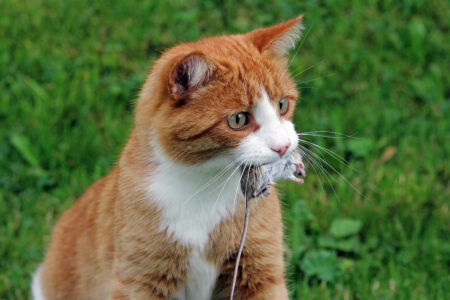
Anticoagulant rodenticides are common poisons used to kill mice, rats, and other rodents. The poison causes life-threatening hemorrhage by interfering with normal blood clotting. Anticoagulant rodenticides are the most frequent cause of poisoning in pets. These poisons are easy to obtain and used anywhere there might be rodents—in homes, garages, farms, cabins, and even wildlife areas. The pet may directly ingest rodent bait, may ingest contaminated food, or may even ingest a rodent that has died from the poison.

MousePoisonCauses and Toxicity

Anticoagulant rodenticide poisons interfere with vitamin K activity in the body. Vitamin K1 is an essential ingredient for the body to make blood clotting factors. Rodent poisons decrease vitamin K1 levels and inhibit the production of clotting factors. Without clotting factors, the body can no longer form clots and uncontrolled life-threatening bleeding may occur.

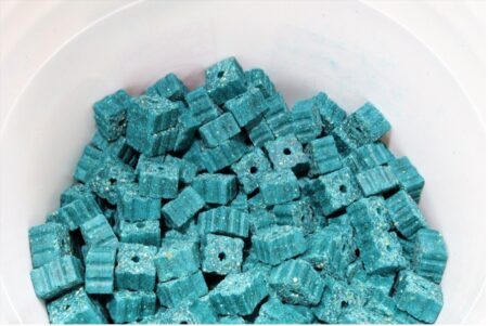
Most of these poisons are bright blue-green pellets, and dogs and cats often confuse them for tasty kibble. Active ingredients in these rodenticides fall into two categories, coumarin-based products (warfarin, coumafuryl, brodifacoum, and bromadiolone) and indanedione-based products (diphacinone, pindone, yalone, and chlorophacinone). Coumarin based products affect vitamin K for days, whereas the effects of indanedione-based products may last for weeks.

Clinical Signs

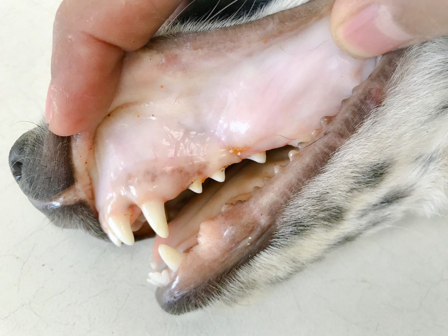
Signs of anticoagulant rodenticide can take several days to develop because vitamin K and clotting factor stores are depleted over time. Clinic signs are related to anemia and bleeding tendencies. External signs of bleeding or bruising are not always present. If the bleeding is internal, weakness and depression may be the only signs noted.

Signs of anticoagulant rodenticides poisoning may include:
• Weakness
• Depression
• Increased respiratory rate
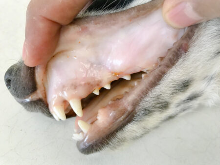
• Pale gums
• Bleeding from nose or mouth
• Lack of appetite
• Enlarged abdomen
• Vomiting
• Bloody or dark tarry stools
• Swelling painful joints
• Bruising
• Neurologic signs

Diagnostic Tests

If ingestion of the rodenticide was witnessed, the diagnosis is straightforward. Not all rodent poisons are anticoagulants, so it is important to identify what active ingredient was ingested.

If ingestion or exposure of the poison was not witnessed, other causes of illness must be ruled out. The veterinarian will start with a physical examination to look for any evidence of external or internal bleeding. Blood tests including complete blood count and serum blood chemistry will likely be recommended for a baseline evaluation. The initial bloodwork may appear to be normal. Special blood tests called coagulation panels will be sent to a reference lab to evaluate the pet’s ability to clot. Additional tests, such as x-rays and an ultrasound, may be recommended to identify internal bleeding in the chest and abdomen.

Treatment

Contact your veterinarian immediately if you suspect your pet has ingested poison. Early decontamination and treatment decrease the risk for serious toxicity. If an anticoagulant rodenticide ingestion occurred within a few hours of seeking treatment, the veterinarian will induce vomiting. Inducing vomiting helps to minimize absorption of the toxin. Activated charcoal may then be administered to further decrease the absorption of anticoagulant rodenticide by the gastrointestinal tract.
The antidote to anticoagulant rodenticide is vitamin K1, a prescription drug. Even if the poison is vomited, treatment with vitamin K1 is usually instituted to ensure patient safety. Vitamin K is given by mouth to increase the clotting factors in the body and prevent bleeding. Oral vitamin K should be administered with food to improve absorption. The duration and dosage of vitamin K1 required depend on the type of toxin ingested. Coumarin products are active for days, whereas indanedione products are active for weeks. If the exact type of toxin is not known, treatment is often given at high doses for 4-6 weeks.

If ingestion of poison occurred days before seeking treatment, and symptoms of bleeding or anemia are present, hospitalization at a 24-hour specialty facility is necessary. In addition to vitamin K1, intravenous fluids and blood or plasma transfusions may be necessary. Oxygen therapy helps support pets with respiratory symptoms. Patients that suffer from internal bleeding may need removal of blood from the chest or abdominal cavity. Repeated blood work to monitor blood clotting times, red blood cell counts, and platelet counts are commonly performed.

Follow-up Care

When anticoagulant rodenticide poisoning occurs, vitamin K1 supplementation must be continued for several weeks. Approximately 48 hours after discontinuing the vitamin K1, blood clotting times will be evaluated to assure no further supplementation is needed. During the early stages of recovery at home, limited activity is recommended to prevent injuries that could cause bleeding. Once vitamin K1 supplementation can be discontinued, the pet can return to regular activities.

Prognosis

Early diagnosis and aggressive treatment are crucial in treating anticoagulant rodenticide poisoning in pets. Pets have their best chances of survival if seen quickly by a veterinarian. Patients that receive prompt treatment including induction of vomiting, administration of activated charcoal, vitamin K therapy, and follow up monitoring have a good prognosis. These pets may make a complete recovery.
Pets that receive delayed treatment after symptoms of anemia and hemorrhage have developed have a more guarded prognosis. These patients may need hospitalized for observation and treatment for 2-6 days, depending on the active ingredient and how much rodent poison was ingested.
Ingestion of rodent poisons can be fatal. Without treatment, the prognosis is grave.

Prevention

Prevention is key! Keep all rodent poisons away from pets. Consider an alternative form of rodent control, such as live traps. If you must use rodent poison keep a detailed record of how much and where the poison was placed. Take a picture of the product and active ingredients in case you need to refer to it later for veterinarian treatment. Never let pets eat unidentified objects, as these may contain poisons that are hard to identify after the fact.

If you suspect your pet ingested an anticoagulant rodenticide, it is important to contact your veterinarian and Pet Poison Helpline, a 24/7 animal poison control center, at 1-800-213-6680 right away to help determine the risk of poisoning to your pet.

Flea Infestation

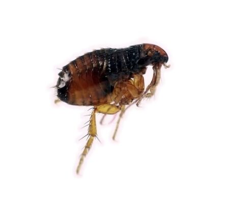
Fleas are insects that consume blood from mammal hosts. Adult fleas are tiny, measuring only 2.5 mm long. Their bodies are shiny and reddish brown in color. Fleas do not have wings, but they can jump long distances. Several species of fleas can infest pets, but the most common one found on cats & dogs is the cat flea or Ctenocephalides felis. Fleas are a public health concern because they can transmit a variety of diseases to pets and people.
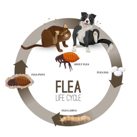
Flea Life Cycle

The flea has a multistage complex life cycle that can be completed in a few weeks:

  • Adults The adult fleas emerge from their cocoons and begin searching for a host. Within minutes of hopping on a pet, the fleas begin feeding on the blood of the host animal.
  • Eggs – Egg production begins within 20-24 hours of female fleas taking their first bloodmeal. The eggs are deposited onto the pet & fall into the environment. Female fleas can produce over 1,000 eggs a month.
  • Larvae – The larvae hatch from the eggs within days. Larvae feed on adult flea feces. The larvae develop outdoors in cool shady areas or may also be found indoors in protected sites in the carpet including under furniture.
  • Pupae – Mature larvae form cocoons that can be found in soil, on vegetation, in carpets and on animal bedding. Adults emerge from the cocoon in one to two weeks depending on the temperature & humidity.

Causes

Cats and dogs can become infected with fleas in their environment or close contact with other animals.

In warmer months, infestations may take root outside your house. Feral and wild animals can bring fleas into your yard.  The adult fleas can wait in cool shady areas for unsuspecting pets to pass by. In colder months fleas can jump from pet to pet by close contact at parks, kennels, play dates, and groomers. Pets will carry the fleas into you home and soon eggs will start shedding into the environment resulting in an infestation.

Symptoms & Diagnosis

Many signs can indicate flea activity:

  • Scratching – Pets infected with fleas repeatedly scratch and over-groom themselves. For pets that are allergic to flea saliva, the bite of even 1 flea can cause intense itching.
  • Irritation – Hair loss, skin redness, and crusty scabs may appear. Pets can cause significant self-trauma from chewing and scratching.
  • Adult fleas – Adult fleas are visible to the naked eye. Fleas can be found on the pet by use of a fine-toothed comb.
  • Flea feces – Flea dirt, the adult flea feces, looks similar to coarse ground black pepper and may be seen in the fur, pet beds, carpets, rugs and other areas where the animal host rests.
  • Anemia – Young animals with severe flea infestations can experience anemia from blood loss. Puppies and kittens may become pale and lethargic with anemia.
  • Tapeworms – A dog or cat may swallow a flea while self-grooming. Ingestion of fleas can infect the host with tapeworms. Owners may see pieces of rice on the stools or in the hair around their rectum. These are tapeworm segments.

A veterinarian can diagnose fleas based on the physical exam findings of skin irritation, and presence of adult fleas or flea dirt.

Treatment

Minor flea infestations that are identified early may be treated by the homeowner in consultation with a veterinarian.  Moderate to severe infestations will take months to control and will require the services of an exterminator.

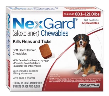
  • Pet treatment – Every pet in the home must be treated. All pets must receive a veterinarian prescribed medication, such as Nexgard, Frontline, or Revolution, administered every month for a minimum of 3 months. Over the counter shampoos and topicals are not recommended, as they often use harsh pesticides with significant side effects and do not break the flea life cycle. Secondary skin irritation and infection may need to be addressed with steroids and antibiotics.
  • Sanitation – Thoroughly clean areas where pets hang out and fleas live. The home should be vacuumed frequently to physically remove flea eggs, larvae, pupae and adults from carpeting, rugs, floors, and furniture. Regularly wash pet bedding in hot water and dry on high heat. The yard should be mowed and raked to remove excess organic debris from flowerbeds, near bushes, and under decks. Feral and wild animals entering the yard should be managed.
  • Home treatment. For moderate to severe infestations, home treatment should begin at the same time as pet treatment. This helps disrupt the flea’s life cycle. A licensed commercial pest control exterminator is needed to determine which products and plan are best for inside the home and in the yard.
  • Follow-up. Fleas have a complex life cycle with multiple stages. To kill fleas, multiple follow-up treatments of the pet and home are needed to eliminate all stages. In the first month, you might see fleas after using recommended products. It takes time for all the adults, larvae, pupae, and eggs to be eliminated from the environment. A minimum of 3 months of treatment for all pets is needed to resolve a flea infestation.

Prognosis

The prognosis for minor flea infestations is excellent if all pets in the home are treated with prescription medications for 3 months and the environment is cleaned regularly. The prognosis for moderate to severe infestations can be guarded. All pets in the home may need to be treated beyond 3 months and a professional exterminator will need to make multiple visits to the property. It is important to remember that fleas can carry numerous diseases; bacteria and parasites that can be transmitted to both pets and people. Diseases of concern include Typhus, Tapeworms, Tularemia, Bubonic Plague, and Bartonellosis. Pets that have been infested with fleas should be monitored closely for additional symptoms that may indicate secondary infections.

Prevention

Flea infections are easy to avoid with regular year-round use of safe and effective prescription medications available from a veterinarian. For dogs, Nexgard and Simparica are once monthly chewable or Frontline and Revolution are once monthly topicals that can be used to prevent fleas and ticks. For cats, Nexgard Combo, Revolution, and Frontline are once a month topicals that help prevent fleas and ticks.

Talk to your veterinarian about the best product to use for your pet. The products you buy from your veterinarian are backed and guaranteed by the manufacturers in cases of reaction or product failure.

For more information visit the Companion Animal Parasite Council www.capcvet.org/guidelines/fleas/

Feline Lower Urinary Tract Disease (FLUTD)

Feline Lower Urinary Tract Disease (FLUTD) is a medical condition characterized by pain and inflammation in the bladder and urethra. Most incidents of FLUTD are caused by Idiopathic Sterile Cystitis. While the condition can be seen in cats of any age, it is most frequently seen in young to middle-aged, overweight cats that get little exercise, use an indoor litter box, have restricted outdoor access, and eat a dry diet.

Symptoms of Feline Urinary Disease

  • Straining to urinate
  • Frequent and prolonged attempts to urinate
  • Crying in pain during urination
  • Blood in the urine
  • Decreased volume of urine
  • Inappropriate urination outside of the litter box
  • Excessive grooming of the belly and genital area
  • Irritability and hiding

Diagnosis of Feline Urinary Disease

A diagnosis of FLUTD can only be made by excluding other causes of lower urinary tract disease.

Possible tests include:

  • Urinalysis to evaluate the composition of the urine including the pH, presence of blood, or crystals.
  • Bacterial culture of a urine sample to identify infections
  • Bloodwork to evaluate general health and screen for diabetes & kidney disease
  • X-rays of the urinary system to identify stones
  • Ultrasound of the bladder and kidneys to diagnose stones, tumors, and polyps

Causes of FLUTD

Over 70% of FLUTD cats have Feline Idiopathic Sterile Cystitis

The majority of cats that develop FLUTD have no obvious underlying medical cause. When no underlying cause can be identified, cats are diagnosed with feline idiopathic cystitis or FIC. This form of disease appears to bear many similarities to a disease in humans called ‘interstitial cystitis’.

FIC is a neurogenic inflammatory condition of the bladder lining triggered and worsened by stress. When a cat’s stress hormones (adrenaline and cortisol) rise, an inflammatory response occurs in the nerves of the bladder lining. The inner lining of the bladder lumen (GAG layer) also begins to deteriorate leaving the nerve endings less protected. This inflammation causes in the classic symptoms of FLUTD. The experience of bladder inflammation causes more stress, resulting in a vicious cycle of rising stress hormones and inflammation.

15% of FLUTD cats have urolithiasis or crystalluria (urinary stones and mineral sediment)

Crystals are mineral deposits that form in urine when the urine pH is abnormal, such as too alkaline or too acidic. Rock-hard collections of crystals/minerals can form urinary stones (uroliths). Crystals and stones can cause many of the common symptoms of FLUTD.

A urinalysis may demonstrate the presence of crystals in the urine. Imaging is needed to make a diagnosis of urinary stones. X-rays are used to visualize stones in the bladder. The treatment depends on the mineral composition of the stones and crystals, but may include a prescription diet or surgical removal.

10% of FLUTD cats have a structural abnormality such as a tumor, polyp, or diverticulum

Structural abnormalities alter how the bladder functions and can inhibit normal bladder emptying. An ultrasound of the bladder and kidneys can identify soft tissue structural abnormalities

Less than 5% of FLUTD cats have Bacterial Urinary Tract Infections

Cat urine normally dicourages bacterial growth. Only cats that are producing abnormal urine are prone to developing lower urinary tract infections. Cats may develop a urinary tract infection if they have very dilute urine from chronic kidney disease, glucose in the urine from diabetes, or blood in the urine from stones or tumors. A positive urine culture can confirm the presence of a bacterial urinary tract infection.

Treatment of FLUTD

The treatment for interstitial cystitis has many components:

Pain Management
All cats suffering from FLUTD experience pain. Buprenorphine is the most common pain medication prescribed. This will last 8-12 hours, and must be given two to three times daily.  This medication is not effective if swallowed; it must be applied to the gums or under the tongue.

Cats may also be prescribed Prazosin and Gabapentin, these medications target the painful urethral muscle spasms and neuropathic pain pathway that contribute to the frequency and straining common in FLUTD. Medications targeted towards inflammation such as steroids or anti-inflammatories may also be used.

Anxiety Medication

The most prescribed antianxiety medications are fluoxetine and amitriptyline. These medications increase the cat’s ability to deal with anxiety and lower the cat’s stress hormones.  Decreasing the levels of stress hormones helps some cats break the cycle of neurogenic inflammation that causes FLUTD symptoms. Anxiety medications are used for cats that have frequent recurrent symptoms of sterile cystitis. 

Antibiotics

Antibiotics will NOT help 95% of cats with FLUTD symptoms. Administration of unnecessary antibiotics may increase FLUTD symptoms due to the stress of having medication forced upon them. An antibiotic should ONLY be prescribed after a positive urine culture or a urinalysis has confirmed a bacterial urinary tract infection. If antibiotics are prescribed, oral medications are most effective (Clavamox or Orbifloxacin).

Decrease the Environmental Stress

If we can point to a recent one-time stressor, such as moving to a new home or upheaval in the daily routine, then once the cat has recovered, there is a good chance that the problem will not recur.

If the cat must contend with daily stress, such as another animal bullying them, recurrent symptoms and long-term therapy with environmental changes are likely to be required. Cats are intelligent and active creatures that can develop anxiety from boredom and lack of stimulation. Increasing exercise and mental stimulation can decrease a cat’s stress. Create a cat friendly environment. Cats need safe spaces such as cat trees to perch on and good hiding places provide an important retreat. Visit www.indoorcat.org  for ideas to create a stress-free environment.

Improve Liter Box Accommodations

The litter box can be a source of stress for many cats. Cats need a clean private bathroom. Most cats prefer a large, open litterbox with litter that is unscented and sandy in texture. Litter boxes need to be cleaned daily; nobody likes a dirty bathroom. Multiple litterboxes are ideal. The recommended number of litter boxes is one per cat plus one extra.  Litter boxes should be distributed in a variety of different locations and levels throughout the house.

Feed Prescription Urinary Cat Food:

Cats with a history of recurrent symptoms of FLUTD or crystalluria should be fed a prescription urinary diet. Controlled research studies have repeatedly demonstrated an 80% lower rate of FLUTD episodes in cats fed the prescription foods compared with cats fed over-the-counter pet store foods.

Prescription urinary diets:

  • Decrease urine concentrations of inflammatory mediators and crystal minerals
  • Increase concentrations of anti-inflammatory nutrients including Omega-3 fatty acids and vitamin E
  • Increase solubility and dissolution of crystals in urine by maintaining proper urine pH
  • Decrease retention of crystals and toxins in the urinary tract by increasing thirst and water intake
  • Provide nutrients that promote emotional wellbeing such as vitamin B and the amino acid tryptophan

The three formulations of prescription food available are Hill’s Science Diet C/D, Purina UR, and Royal Canin SO. All of these foods work well to prevent the incidence of FLUTD. The cat’s personal taste preferences often determine which food is used long term.

Increase Water Intake

Cats evolved as desert creatures; they do not instinctively drink enough water.  Highly concentrated urine is more likely to irritate the delicate bladder lining and minerals are more likely to precipitate out to form crystals or stones. The toxic substances in concentrated urine will contribute to FLUTD.

Ways to increase water intake:

  • Feed more canned/wet food. Cats should be fed canned food daily and have less access to dry kibble.
  • Improve the drinking water source. Distilled or bottled water may be better accepted.
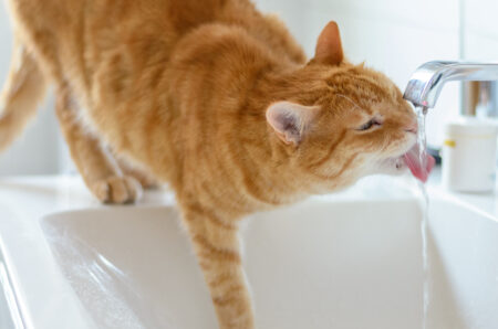
  • A pet drinking fountain or a dripping faucet may appeal to some cats that enjoy running water.
  • Water bowls should be made of porcelain or glass, cleaned daily, and be wide and shallow.

Hypothyroidism in Dogs

The thyroid gland helps to regulate the body’s metabolism by producing hormones. Hypothyroidism is an underactive thyroid gland. In dogs, hypothyroidism is commonly diagnosed between the ages of four to ten years.  Hypothyroidism occurs more commonly in large breed dogs.

Causes

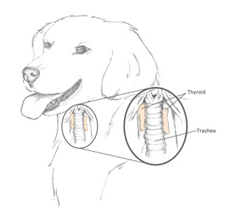
The thyroid gland is a paired lobed gland located in the throat area on either side of the trachea.  Hypothyroidism in dogs is thought to be an immune-mediated disease called lymphocytic thyroiditis. The immune system attacks the cells in the thyroid gland causing inflammation and atrophy of the gland. Over time the thyroid gland’s activity decreases with a subsequent decreased production in the thyroxine hormone. It is unclear why this condition occurs; however, it is a heritable trait meaning that genetics likely plays a role in this condition.

Symptoms

  • Loss or thinning of hair
  • Dull and dry hair coat
  • Thickening of the skin and increased skin pigment
  • Lack of energy
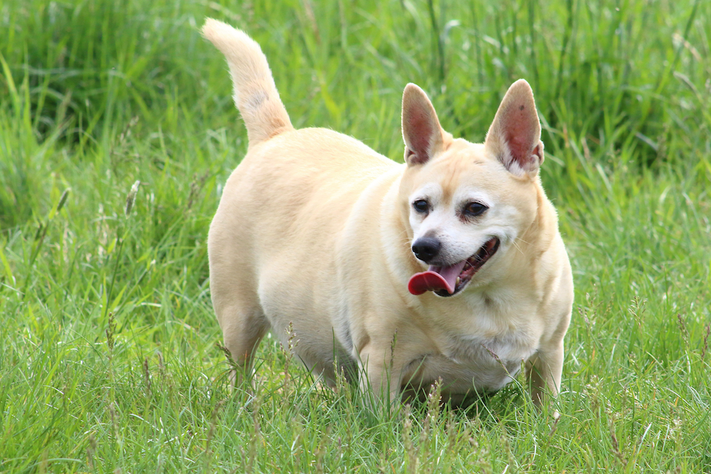
  • Weight gain
  • Reduced ability to tolerate the cold
  • Neurologic changes including dragging of feet and a head tilt

Diagnosis

There are several types of blood tests used to diagnose and monitor hypothyroidism including Total T4, free T4, and TSH. Thyroxine (T4) is the primary hormone produced by the thyroid gland. T4 circulates in the blood in two forms; one form of the hormone is bound, or attached to proteins in the blood, while the other form circulates freely within the blood stream. TSH (Thyroid Stimulating Hormone) is produced by the pituitary gland to regulate the thyroid gland’s activity.

  • Total T4 (thyroxine) – Total T4 level can be measured by a blood test to evaluate both forms of the thyroxine hormone. If the total T4 concentration is below the normal range, and the dog has supportive clinical signs, then hypothyroidism is likely. Total T4 is commonly included on senior screening blood work to help identify subclinical patients.
  • Free T4 (free thyroxine) – This test measures the amount of the free thyroxine hormone in a blood sample. Unfortunately, there are conditions other than hypothyroidism that may cause the total T4 value to be reduced. These conditions include the presence of another illness or the administration of some drugs. Free T4 is less affected by the presence of other illnesses or drugs. If free T4 is below normal range then hypothyroidism is confirmed.
  • TSH (Thyroid Stimulating Hormone) – TSH levels can be measured by a blood test. In hypothyroid dogs, the concentration of TSH may increase as the pituitary gland tries to stimulate an underactive thyroid gland to increase hormone production. If the total T4 and free T4 levels have not provided a clear diagnosis, an elevated TSH can help confirm the presence of hypothyroidism.

Treatment

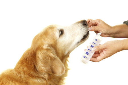
Hypothyroidism is treatable but not curable. The recommended treatment is administration of an oral synthetic thyroid hormone replacement called levothyroxine. Thyro-Tabs, an FDA approved formulation of levothyroxine comes in a pill is available. Cost effective generic medications are also available. The medication is normally given once or twice a day.  The dose and frequency administered will be determined by your veterinarian. Levothyroxine is generally well tolerated with minimal side effects. Treatment of hypothyroidism is life long, meaning this drug must be given for the rest of the dog’s life.

Follow-up

Canine patients with hypothyroidism will need regular progress exams and diagnostic tests performed by a veterinarian. Once the dog is tested and put on medication, they will have to be rechecked one month later to verify the levothyroxine dose is effective.  Your pet will have to be on thyroid medication for the rest of its life.  Routine thyroid testing should be done on a yearly basis or if symptoms change.

Prognosis

With treatment, the prognosis for hypothyroid dogs is good. Hypothyroidism cannot be cured, but most symptoms will improve with consistent administration of medication. Pets that consistently receive their medication and are monitored regularly by their veterinary have a normal life expectancy. Treatment and control of the hypothyroid pet generally provides very rewarding results for both the patient and the owner.


Kidney Disease in Pets

Kidney2

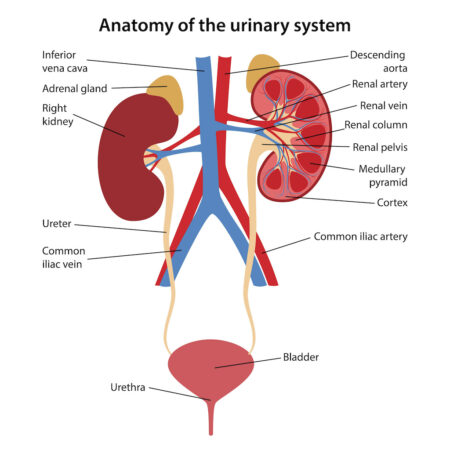
Kidney4The kidneys are remarkable and complex organs that serves many essential functions.

Essential kidney functions:

  • Remove protein wastes and toxins from the body.
  • Balance the body’s fluids.
  • Keep electrolytes at optimal levels.
  • Control pH levels of the blood
  • Release hormones that regulate blood pressure.
  • Maintain the production of red blood cells.

Kidney disease occurs when one or more of these essential functions are compromised or reduced. When kidney function is impaired, body systems become imbalanced. When function is significantly impaired, waste levels can become toxic. Kidney disease can progress to kidney failure.

Causes of Kidney Disease

Infections – Bacterial infections that can cause kidney failure in dogs include Leptospirosis and Lyme disease. Leptospirosis is contacted through urine contaminated drinking water. Lyme disease is acquired through the bite of a deer tick. Both cats in dogs can also acquire ascending infections from the lower urinary system. Untreated bladder infections can cause kidney infections.

Toxicity – The kidney is susceptible to adverse toxicity reactions due to its role in filtering the blood. Common toxins that can cause acute kidney failure include grapes/raisins (dogs), antifreeze, rodenticides, Ibuprofen, and lilies (cats).

Cancers – Renal carcinoma is the most common primary renal tumor in dogs and cats. These tumors have aggressive malignancies with the potential for widespread metastasis. Cancers, including lymphoma, can also originate in other parts of the body and metastasize to the kidneys.

Congenital – Inherited malformation of the kidneys can present in pets under a year of age. The most common condition is polycystic kidney disease in Persian and domestic long-haired cats. The condition causes multiple fluid filled cysts to form within the functional part of the kidney.

Immune Mediated – Glomerulonephritis occurs when a mixture of antibodies and pathogens are filtered out of the bloodstream.  When these compounds are trapped, the body responds by activating its immune defenses, resulting in damage to the kidney tissue.

Urinary Obstruction – Stones may form in the kidneys or bladder. If the stones move from the kidneys into the ureters or from the blader into the urethra, an obstruction that impedes the flow of urine may result. Restricted urine flow can cause a secondary kidney failure.

Chronic Disease – Many senior pets experience a gradual loss of kidney function over time. For older animal populations kidney disease affects up to 10% of dogs and 35% of cats. The exact inciting incident or cause is often not identified in these patients.

Kidney2Clinical Signs

Kidney disease may be detected on routine screening of blood and urine prior to the onset of clinical signs.

A few signs to watch for include:

  • Drinking more water
  • Increase in urine frequency or volume
  • Loss of interest in playing
  • Decrease in appetite
  • Vomiting
  • Weight & muscle loss
  • Poor haircoat
  • Bad breath

Unfortunately, kidney disease typically goes undetected and clinical signs do not appear until the kidneys are functioning at 33% or less of original capacity.

Diagnostic Tests

Diagnosis of kidney disease may require a variety of tests including blood work, urine tests, imaging, and blood pressure.

Blood Tests

Biochemistry Panel – BUN, CREA, and SDMA are measured by a blood chemistry test. Elevations (azotemia) can indicate the kidneys’ declining ability to filter the blood.

Electrolytes & Minerals – Levels of blood electrolytes including potassium, sodium, and chloride may be abnormal. Minerals such as calcium and phosphorous may also be elevated.

CBC – Evaluates levels of red blood cells to look for anemia and white blood cells to identify infection.

Titers – Blood tests for infection including Lyme Disease and Leptospirosis in dogs.

Kidney1Urine Tests

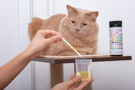
Urinalysis – Analysis of urine by physical, chemical, and microscopic means to test for the presence of disease.

Urine Culture & Susceptibility – The best test to diagnose a urinary tract infection is a culture to grow and identify bacteria present in the urinary tract. The bacteria are then further tested to determine the best antibiotic to use in treatment of the infection.

Urine Protein/Creatinine Ratio – A simple test that measures how much protein is being lost through the kidneys.

Imaging

X-Rays – Radiographs help to assess the size and shape of the kidneys. Kidney enlargement, atrophy or shrinkage, and abnormal shapes or asymmetry may indicate disease. The presence of masses or tumors that change the shape of the kidney can sometimes be visualized using radiographs. Kidney and bladder stones may also be identified on x-rays.

Ultrasound – Ultrasonography is the best diagnostic imaging modality to evaluate the internal structure of the kidneys. Ultrasound reveals any changes in the internal anatomy of the kidney, including abnormal growths, stones, or blood flow.

Blood Pressure – Monitoring blood pressure in kidney patients is important.  High blood pressure (hypertension) is a very common symptom associated with chronic kidney disease. Hypertension causes further deterioration of kidney function.

Classifications and staging

Kidney disease in pets can be classified as acute or chronic. Acute kidney failure happens quickly over several days, and is often a result of sudden kidney injury caused by a specific event such as toxin, obstruction, or infection. Chronic kidney failure happens slowly over months to years. Chronic kidney disease is more prevalent in senior pets and progresses with age.


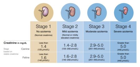
Kidney disease is staged based upon results of diagnostic tests including the level of nitrogen waste build up (azotemia), amount of protein in the urine, and blood pressure. The stage of disease helps to guide therapy and to determine prognosis.  

Treatment Options

Although there is no definitive cure for kidney disease, treatment can often improve and prolong the lives of pets with this disease. Therapy is geared toward minimizing the buildup of toxic waste, maintaining hydration, addressing disturbances in electrolytes, supporting appropriate nutrition, controlling blood pressure, and slowing the progression of kidney disease.

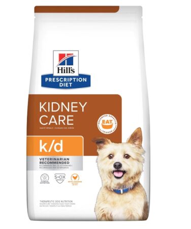
Kidney5Nutritional Therapy – Prescription therapeutic foods, such as Science Diet KD or Purina NF, are formulated with low levels of phosphorous and moderate amounts of high quality protein. These diets are highly palatable and calorie dense to support debilitated animals. Prescription renal diets have domonstrated in studies to prolong survival time, slow disease progression, and improved quality of life. 
Hydration – Kidney disease patients are prone to dehydration due to body’s inability to conserve water. Patients must always have free access to water. Severely dehydrated patients may need to be hospitalized for IV fluids. Moderate chronic dehydration may be treated with subcutaneous fluids, which are fluid injections under the skin. Feeding of canned food can naturally increase daily water intake for pets experiencing mild dehydration.

Supplements – Additional potassium supplementation may be needed in some cats with chronic kidney disease. Control of blood phosphorus levels is also necessary. If dietary changes alone do not accomplish this, drugs to bind the phosphorus in the food can be given with each meal.

Detox – With acute toxin exposures, a veterinarian can take steps to minimize absorption of toxins. Depending on the situation, the veterinarian may induce vomiting or pump a pet’s stomach, administer activated charcoal, and administer an appropriate antitoxin.

Medications – Patients with acute infectious causes of kidney disease should be treated with broad-spectrum antibiotics. Ideally, the antibiotic choice should be based on test results including titer and bacterial culture. Pets that have poor appetites or nausea may benefit from antacid and anti-nausea medications, such as famotidine and Cerenia. If hypertension and proteinuria is present, a variety of drugs, such as Ace inhibitors, can be used to control the high blood pressure.

Prognosis

The prognosis for pets with kidney disease varies depending on the inciting cause, timeliness of diagnosis, and therapy provided.

Early diagnosis and intervention are important for successful treatment of dogs and cats with kidney disease.  Pets that have a delayed diagnosis will have a poorer prognosis and may only live weeks to months. Patients that are diagnosed in the early stages of disease can live several years with conscientious management of their disease by the veterinarian and pet owner. On average cats tend to live longer than dogs with kidney disease.

In most cases, kidney disease is a progressive disease that worsens over time, but the rate of progression is highly variable. Regular follow up exams and lab tests help the veterinarian determine the progression of an individual animal’s kidney disease and make tailored changes in medical management.

Successful treatment of kidney disease delays progression, provides greater survival times, and increases patient quality of life.

Chocolate Toxicity

chocolate toxicity

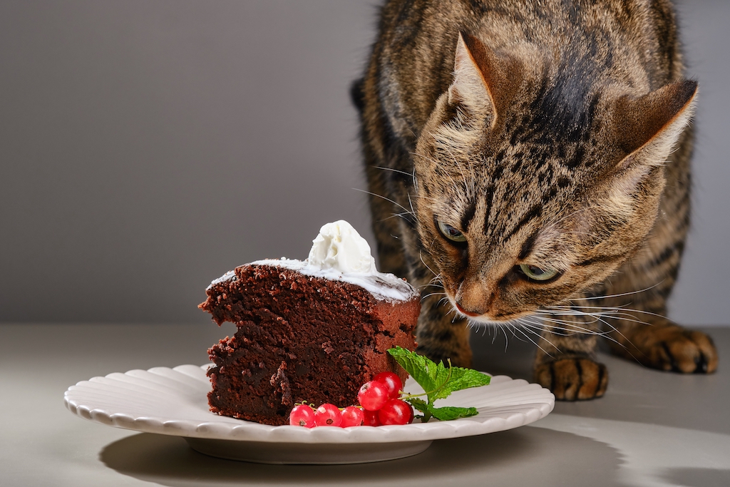
Ingestion of chocolate can be a deadly mistake for dogs and cats. Chocolate (cacao) contains the substances caffeine and theobromine.  These substances can have detrimental effects on the GI system, central nervous system, heart, and kidneys of pets. In dogs and cats the ‘half-life’ (length of time it takes for 50% of the substance to break down in the body) for theobromine is over 17 hours.  This means that after 17 hours, half of the theobromine ingested is still in their systems. Humans can metabolize caffeine and theobromine more efficiently than pets can, which is why chocolate is not toxic to humans.

How much chocolate is considered toxic?

chocolate toxicityNo amount of chocolate is safe for an animal to ingest. The effects of chocolate consumption in pets depends on type of chocolate, quantity consumed, and the size (body weight) of the pet. Some types of chocolate contain higher amounts of caffeine and theobromine.

Reported combined caffeine & theobromine content in different chocolates:

  • Dry cocoa powder contains 800 mg/oz
  • Unsweetened baker’s chocolate contains 450 mg/oz
  • Cocoa Bean hulls (mulch) contains 255 mg/oz
  • Semisweet dark chocolate contains 150-160 mg/oz
  • Milk Chocolate contains 64 mg/oz
  • White chocolate contains an insignificant amount.

The most toxic, cocoa or baker’s chocolate, causes a life-threatening emergency when ingested. The moderately toxic dark chocolates can lead to hospitalization and serious illness in pets when amounts as small as one-tenth of an ounce per pound of body weight are consumed. Even pets that devour less than a half ounce of milk chocolate per pound of body weight are at risk of developing clinical signs. 

Online chocolate toxicity calculator can be helpful for pet owners and veterinarians to calculate the potential toxicity of chocolate ingestion.

https://www.petmd.com/dog/chocolate-toxicity

Symptoms of Chocolate Ingestion

Clinical signs of chocolate toxicosis usually occur within 6–12 hours of ingestion.

Initial signs may include:

  • Increased thirst
  • Vomiting
  • Diarrhea
  • Restlessness

Signs may progress to:

  • Hyperactivity
  • Increased urination
  • Incoordination or trouble walking
  • Fever
  • Twitches & tremors
  • Rigidity & seizures
  • Racing heart progressing to abnormal rhythms

chocolate toxicityTreatment of Chocolate Toxicity

If you suspect your pet has ingested chocolate, you should immediately contact your veterinarian.

If ingestion has occurred within the past 2 hours, the first step is to recover as much chocolate from the patient as possible by inducing vomiting. For many patients, removing the undigested chocolate converts the toxicity from a life-threatening poisoning to just an upset stomach that is easily managed.  Absorbents, such as activated charcoal, are administered by mouth to bind the chocolate and prevent absorption of theobromine and caffeine through the gut. The patient may be administered SQ fluids to help flush the system. Pets may be treated on an out patient basis if minor quantities of chocolate have been ingested and treatment is started before clinical signs develop.

If a pet has consumed larger quantities of chocolate, has developed clinical symptoms, or treatment has been delayed then hospitalization may be required.  Hospitalized patients receive IV fluids to help flush the kidneys and frequent trips out to go potty are needed to prevent re-absorption of the toxins.  Laboratory blood testing may be required to monitor the kidneys. ECG will be used to monitor the heart for changes in rate and rhythm. A variety of medications are used to help treat symptoms such as seizures and arrythmias. Patients may need intensive care for 24 to 48 hours.

Prognosis
The prognosis after chocolate ingestion can be variable depending on the type and quantity of chocolate consumed, and the timing of treatment.

The prognosis is favorable if decontamination, including induction of vomiting and administration of activated charcoal occurs within 2 hours of ingestion and before clinical signs began, the prognosis is favorable. Patients that receive prompt treatment may be treated as outpatients and their symptoms may not progress beyond mild GI upset.

If treatment is delayed past 4 hours of ingestion, and mild clinical signs have started, the pet will need to be hospitalized for 12 to 36 hours for monitoring and supportive care. Pets that have developed symptoms of toxicity have a more guarded prognosis. If symptoms progress to seizures and cardiac arrhythmias the prognosis is poor, and the toxicity may be fatal.

In the days following chocolate ingestion, patients should be monitored carefully. The high fat content of chocolate products may trigger secondary pancreatitis in some patients.


Corneal Ulceration

Description
A corneal ulcer occurs when the protective surface layer of the eye’s cornea is lost or damaged. Corneal ulcers range from superficial abrasions and small circular lesions to deep craters. The deeper layers of the cornea are exposed and become prone to infection and injury. These deeper layers contain many nerves, and irritation of these nerves is very painful. Ulcers may be complicated by secondary bacterial infections.

Causes
There are many causes of corneal ulceration, including:
• Trauma from cat scratches or other foreign objects
• Abnormalities of the eyelids including entropion (eyelids rolled inward) or abnormal ingrown eyelashes
• Decreased blinking due to neurologic problems or from sedation and general anesthesia
• Exposure to irritants such as chemicals, soaps, or smoke
• Infections with bacteria, viruses, or fungal agents
• Chronic dry eyes from poor tear production
• Corneal infiltrates such as calcium, cholesterol, or edema
• Brachycephalic breeds of dogs or cats with flat faces and prominent eyes are very prone to corneal ulcers because their eyes protrude beyond the eyelids.

Clinical Signs
Pain is a hallmark sign of corneal injuries and ulceration. A pet with a painful eye may have increased tearing, squinting, blinking, and pawing at the eye. The animal may also be quiet and withdrawn. The eye is usually red and thick eye discharge may develop with infection. The cornea may be cloudy or have visible irregularities. Other signs, such as swelling of the eyelid, inward rolling of the eyelid, protrusion of the third eyelid, or bruising around the head, may occur depending on the cause of the ulceration. Signs of an upper respiratory tract infection may be noted in cases of herpes virus infection in cats or distemper virus infection in dogs.

Diagnostic Tests
A veterinarian diagnosis a corneal ulcer by performing an ophthalmic exam.

Fluorescein staining of the cornea is used to confirm the presence of a corneal ulcer. The stain adheres to deeper corneal layers that are exposed when the corneal surface is damaged. The damaged area if cornea is visible as apple green under blue light. Other ocular tests such as a Schirmer tear test, testing of reflexes, examination of the eyelids and interior of the eye, and glaucoma testing may also be indicated. If a severe bacterial infection is suspected a culture may need to be submitted.

TREATMENT AND FOLLOW-UP

Treatment Options
Most corneal ulcers in dogs and cats can be treated on an outpatient basis by a general veterinarian.

Medications and treatments prescribed for corneal ulcers:
• Topical antibiotics drops or ointment administered directly to the eye to treat active infections and/or to prevent infections.
• If the ulcer is deep and there is concern about infection within the eye, then oral antibiotics may also be prescribed.
• Topical atropine (pupil dilator) may be administered as an ointment or drop directly to the eye.
• Oral nonsteroidal anti-inflammatory drugs may be considered for marked inflammation and discomfort.
• Administration of the pet’s own blood serum as eye drops to provide nutrients to encourage healing.
• Elizabethan cone collar to prevent self-trauma.
• Debridement of the ulcer with cotton swab to remove dead tissue

Referral to a veterinary ophthalmologist may be recommended if the ulcer is deep or not healing. An ophthalmologist can offer application of soft contact bandage lenses or placement of a third eyelid flap to protect the cornea. An ophthalmologist can also offer treatments for underlying conditions such as entropion correction, removal of foreign materials, or abnormal eye lashes.

Follow-up Care
Recheck exams are needed to assess healing and response to treatment. Visit frequency can range from every 24-48 hours to every 7-14 days, depending on the severity of the corneal ulcer. Fluorescein staining of the cornea is performed at most visits to highlight the ulcer and to determine when it has healed. Brachycephalic breeds such as boxers, bull dogs, and Persians may need more intensive follow-up.

Prognosis
Most superficial corneal ulcers and abrasions caused by trauma respond to treatment quickly and heal with minimal scarring. Pets that have underlying chronic conditions such as entropion or ingrown eye lashes may need to have minor procedures to correct the abnormality. Chronic conditions, such as dry eye, may need lifelong treatment with eye drops to prevent future corneal ulcers. Deep ulcers and ulcers that are infected are more difficult to treat and may leave the cornea scarred and pigmentated resulting in decreased vision. In rare instances corneal ulcers that that do not receive treatment and become infected can result in perforations of the eye and loss of vision, and in severe cases even loss of the eye.